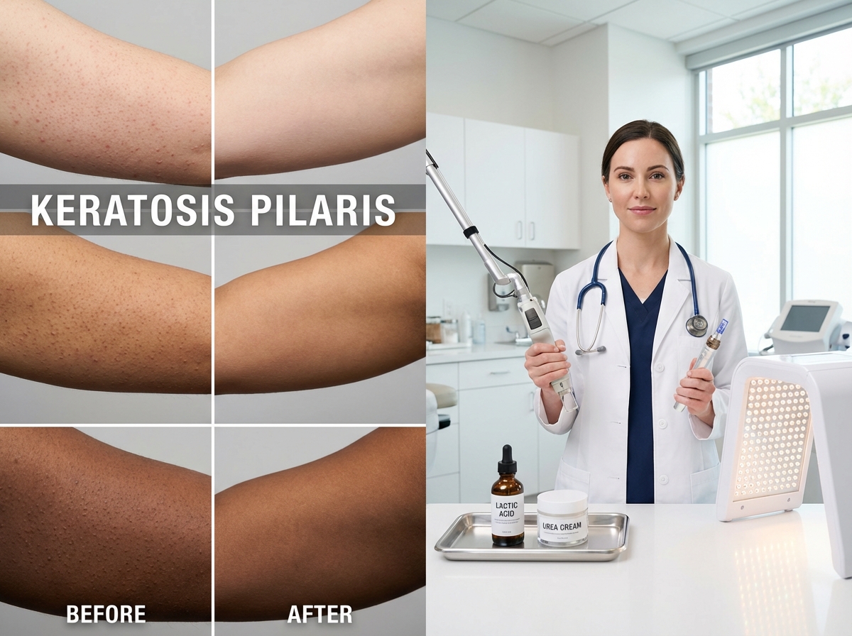
Keratosis pilaris is a common follicular keratinization disorder that causes rough bumps and sometimes redness on the arms, thighs, and cheeks. This article explores noninvasive rejuvenation options—lasers, microneedling, chemical peels, RF, and LED—and how to combine them with topical exfoliation for safer, realistic improvement, outlining indications, downtime, expected results, and safety advice.
Understanding Keratosis Pilaris and Noninvasive Rejuvenation
Keratosis pilaris (KP), often colloquially called “chicken skin,” is one of the most common skin conditions seen in dermatology clinics. Despite its prevalence, affecting up to 80% of adolescents and 40% of adults, it remains a source of significant cosmetic concern. Understanding its underlying cause is the first step toward effective management. At its core, KP is a disorder of follicular keratinization. The body produces an excess of keratin, a protective skin protein, which accumulates and forms hard plugs within individual hair follicles. These plugs create the characteristic small, rough bumps.
The condition typically appears on the outer-upper arms, thighs, buttocks, and sometimes the cheeks in children, giving the skin a sandpaper-like texture. While the bumps are the primary feature, they are often accompanied by varying degrees of redness, known as keratosis pilaris rubra, or post-inflammatory hyperpigmentation, which leaves behind small, dark spots, especially in individuals with darker skin tones. The natural history of KP varies; for many, it appears during childhood or puberty and may gradually improve with age. For others, it persists well into adulthood, often fluctuating with seasonal changes and worsening in dry winter months.
While medically benign, the psychosocial impact of keratosis pilaris is significant. The rough texture and visible bumps can lead to self-consciousness, causing individuals to avoid clothing like sleeveless tops or shorts. This desire for smoother, clearer skin drives most patients to seek treatment. The goal of any management plan is not a permanent cure—as KP is a chronic genetic condition—but rather significant improvement and long-term control. Primary objectives include smoothing the skin’s texture by reducing keratin plugs, decreasing visible bumps, lessening associated redness or discoloration, and establishing a consistent maintenance routine to prevent recurrence.
Treatment follows a hierarchical approach, starting with accessible options and escalating to advanced procedures. The foundation of any effective regimen is consistent at-home care.
- Topical Keratolytics and Emollients: These are first-line treatments. Keratolytics, such as lotions containing lactic acid, salicylic acid, or urea, work by dissolving keratin plugs and promoting exfoliation. Emollients and rich moisturizers soften the skin and reduce dryness, making bumps less noticeable.
For many with mild KP, this foundational care is sufficient. However, for persistent or severe presentations, in-office procedures offer a significant boost by targeting specific aspects of the condition.
- Chemical Peels and Microdermabrasion: These provide more intensive exfoliation than topical products, removing the top layer of dead skin cells to unblock follicles and improve texture.
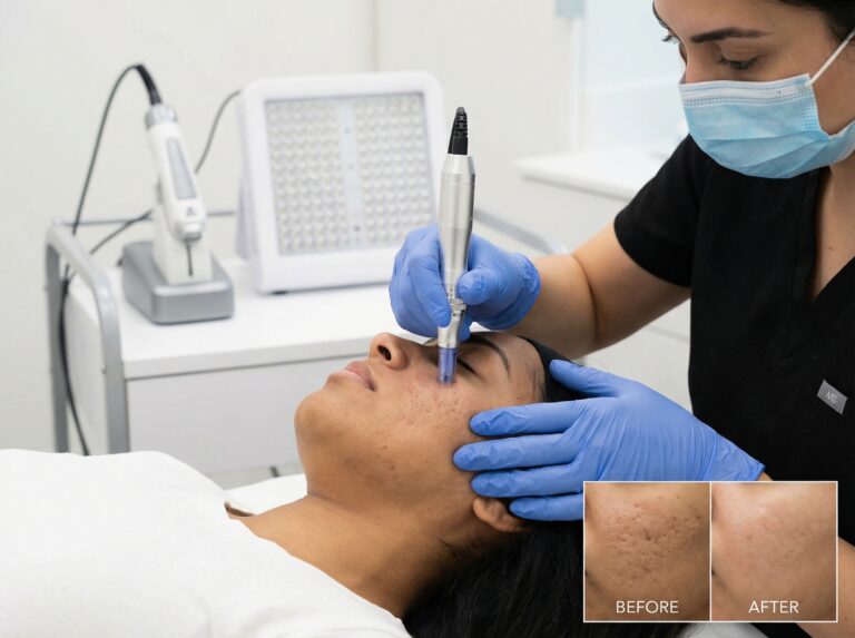
- Microneedling: This modality creates micro-injuries to stimulate collagen production and skin remodeling, effectively improving the rough texture associated with long-standing KP.
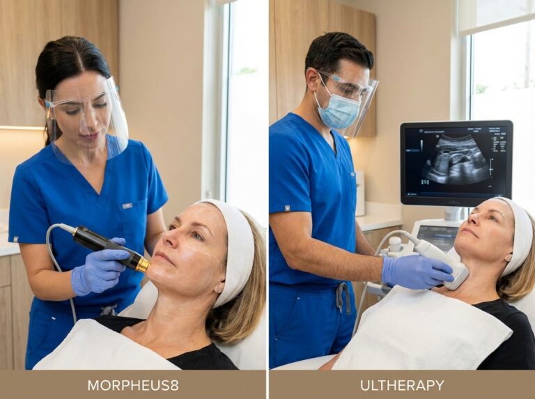
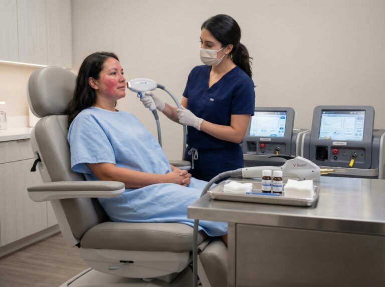
- Vascular and Fractional Lasers: Lasers offer a targeted approach. Vascular lasers, such as the pulsed dye laser (PDL), target hemoglobin to reduce persistent redness (erythema). Fractional lasers create microscopic treatment zones to resurface the skin, dislodging keratin plugs and smoothing texture.
- Hair Removal Lasers: Since KP is a disorder of the hair follicle, reducing the hair itself can improve the condition. Lasers designed for hair removal decrease the prominence of the follicle and associated plugging.
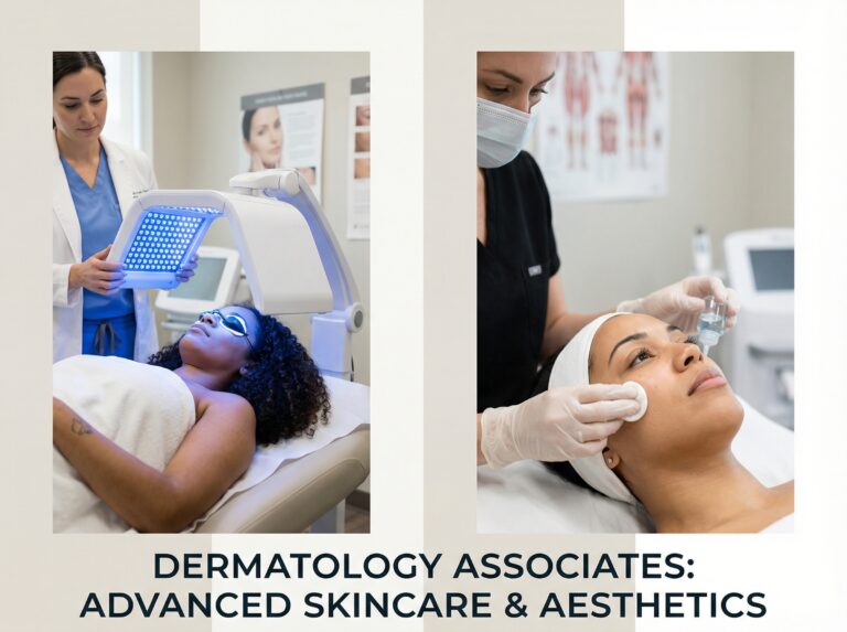
- Radiofrequency (RF) and LED Therapy: RF aids in skin remodeling, while LED therapy (particularly red light) may help reduce inflammation and redness.
Patient selection is key. Individuals with mild textural irregularities are often candidates for topical therapy alone. Escalation to in-office procedures is warranted when bumps are stubborn, texture is significantly rough, or redness is the primary concern. For instance, a patient bothered mainly by redness would benefit more from a vascular laser than microdermabrasion.
It is important to approach KP treatment with realistic expectations grounded in clinical evidence. While these modalities show promise in practice and small studies, high-quality, large-scale randomized controlled trials are limited. Improvement is possible, but complete and permanent clearing is unlikely. Success is defined as achieving smoother, less inflamed skin through a combination of professional treatments and diligent home care. For a clinical overview, refer to the NCBI Bookshelf article Keratosis Pilaris – StatPearls.
Targeted Laser and Exfoliation Strategies for Keratosis Pilaris
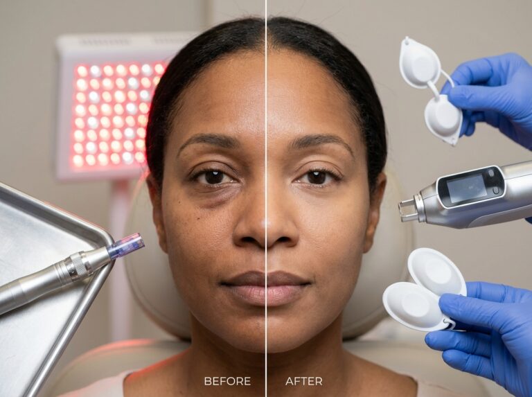
When topical treatments alone do not provide the desired smoothness, in-office procedures can be layered into a treatment plan. These strategies target the different components of KP: keratin plugs, rough texture, redness, and follicular prominence. Evidence suggests that combining modalities often yields better results than any single approach.
Lasers for Targeted Improvement
Lasers offer a precise way to address specific features of KP. The choice of device depends on whether the primary concern is texture, redness, or the hair follicle itself.
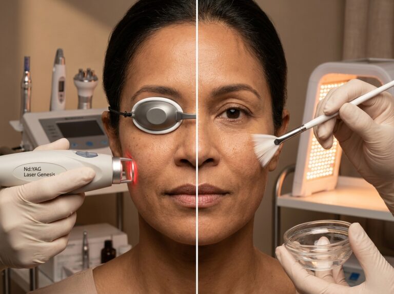
- For Rough Texture: Non-ablative fractional lasers, such as the Erbium Glass (1540–1550 nm) and fractional Erbium YAG, are used to improve skin texture. These devices create microscopic columns of thermal injury deep within the skin, leaving the surrounding tissue intact. This stimulates collagen remodeling and normalizes the follicular environment, reducing keratin plugging over time.
- For Persistent Redness: When KP presents with significant erythema, vascular lasers are the gold standard. The Pulsed Dye Laser (PDL) and other 585–595 nm wavelength devices target hemoglobin within the small blood vessels surrounding hair follicles. The laser energy collapses these vessels, reducing the overall red appearance. Intense Pulsed Light (IPL) with appropriate filters can also be effective for diffuse redness.
- For Follicular Prominence: In cases where KP is associated with dark, coarse hairs trapped within bumps, hair removal lasers are beneficial. Long-pulsed Nd:YAG (1064 nm), Alexandrite (755 nm), and Diode (810 nm) devices target pigment in the hair follicle. By reducing or eliminating the hair, these treatments decrease follicular plugging and inflammation.
Treatment Protocol: A typical course involves 3 to 6 sessions spaced 4 to 8 weeks apart. Improvements are often modest and gradual, with full textural benefits appearing 3 to 6 months after treatment begins. Maintenance sessions are typically required to sustain results.
Advanced Exfoliation Strategies
Building on at-home care, professional exfoliation intensifies the breakdown of keratin plugs.
Topical Keratolytics and Retinoids
Consistent home care is the backbone of management. Over-the-counter products containing lactic acid (10-15%), urea (10-20%), or low-concentration salicylic acid are starting points. For resistant cases, prescription-strength formulations (e.g., urea up to 40%) are powerfully keratolytic. Retinoids like tretinoin and adapalene accelerate skin cell turnover, preventing the buildup of dead cells that form plugs.
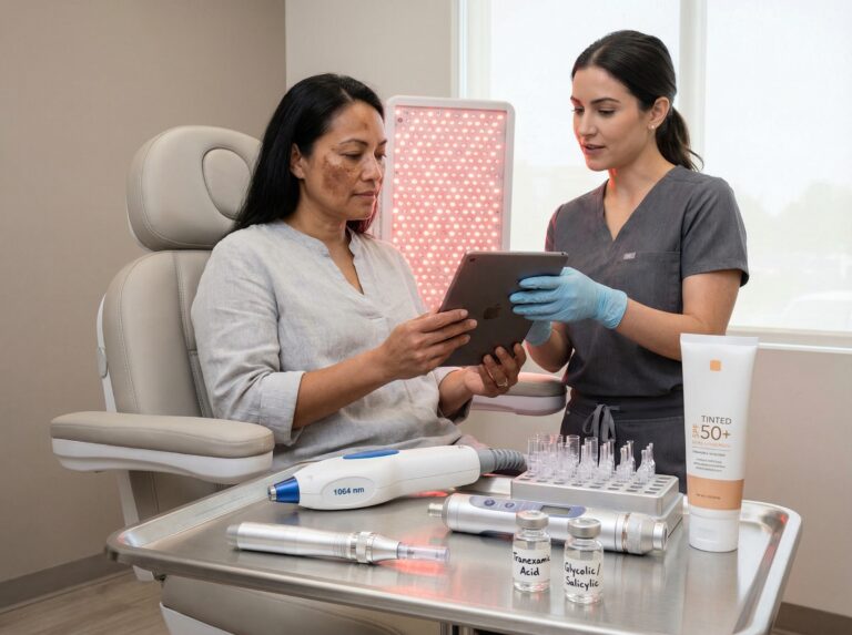
In-Office Chemical Peels
Professional peels offer deeper, controlled exfoliation. For KP, superficial peels are preferred to minimize risks.
- Alpha-Hydroxy Acids (AHAs): Glycolic and lactic acid peels (typically 30-70%) dissolve bonds between dead skin cells.
- Beta-Hydroxy Acids (BHAs): Salicylic acid peels (20-30%) are particularly useful as they are oil-soluble, penetrating the follicle to dissolve plugs from within.
- Light TCA Peels: A very light trichloroacetic acid (TCA) peel (10-15%) may be used for texture, though caution is required regarding pigmentation changes.
Peels are often performed in a series of 4 to 6 treatments every 2 to 4 weeks. Downtime is minimal, usually involving mild redness and flaking for 2 to 7 days.
Microneedling and Radiofrequency (RF) Microneedling
Microneedling uses fine needles to create controlled micro-injuries, triggering a wound-healing response that stimulates collagen. RF microneedling enhances this by delivering radiofrequency energy to generate heat in the dermis for robust tissue tightening. These treatments are synergistic with lasers and peels, offering a mechanical method to break down rough texture. Downtime typically involves 1 to 5 days of redness, mild swelling, and a temporary “sandpaper” texture.
Combining Therapies and Preparation
A multi-pronged approach is often most effective. A clinician might design a protocol involving pre-conditioning the skin with topical retinoids or keratolytics for several weeks to thin the outer layer, making subsequent procedures more effective.
Preparation Guidelines:
- Discontinue Irritants: Stop retinoids, AHAs, and BHAs 3 to 7 days before professional treatment to avoid excessive irritation.
- Sun Avoidance: Avoid direct sun exposure and tanning beds for at least two weeks prior to treatment.
- Spacing: To avoid compromising the skin barrier, wait at least 2 weeks between a chemical peel and a laser or microneedling treatment on the same area. For example, a patient might alternate monthly between a salicylic acid peel and a vascular laser session.
Safety, Costs, and Considerations
Safety is paramount, particularly for skin of color (Fitzpatrick types IV-VI), where the risk of post-inflammatory hyperpigmentation (PIH) is higher.
Safety Protocols for Darker Skin:
- Wavelength Selection: Clinicians should utilize longer wavelengths, such as the 1064 nm Nd:YAG, which bypasses epidermal melanin to target deeper structures safely.
- Conservative Settings: Lower energy and longer pulse durations are essential to prevent burns or pigment changes.
- Test Spots: A patch test in a less visible area is mandatory to observe the skin’s reaction before full treatment.
- Pre-treatment: Pre-treating with depigmenting agents like hydroquinone may be recommended.
Contraindications: Procedures should be postponed if there is an active infection in the treatment area. Recent use of oral isotretinoin typically requires a waiting period of 6 months due to potential impairment of wound healing.
Costs and Downtime:
Costs vary by region and provider. Chemical peels are generally the most affordable ($150-$400 per session), followed by microneedling ($200-$700), with lasers being the most expensive ($300-$800+). Downtime ranges from a day of mild redness for a light peel to several days of redness and swelling for fractional laser treatment.
Sample Stepwise Treatment Plans
- Mild KP: Home Care: Daily use of lotion with 12% lactic acid or 20% urea. Procedures: Optional series of 3-4 light salicylic acid 20% peels every 4 weeks. Maintenance: Continued home care.
- Moderate KP (Texture and Redness): Home Care: Alternating a prescription retinoid (e.g., adapalene 0.1%) at night with a strong keratolytic moisturizer in the morning. Procedures: A series of 4 PDL sessions for redness, alternating monthly with 4 RF microneedling sessions for texture. Maintenance: Consistent home care and one maintenance procedure every 4-6 months.
- Recalcitrant KP: Home Care: As above, potentially with 40% urea cream applied under occlusion. Procedures: Start with 4-6 laser hair removal sessions (e.g., Nd:YAG) to address the follicular component. Follow with 3-4 non-ablative fractional laser treatments for residual texture. Maintenance: Diligent home care and annual touch-up sessions.
Conclusions and Practical Recommendations
Navigating the treatment landscape for Keratosis Pilaris can feel overwhelming, but the path to smoother skin is built on a logical progression of care. At its core, KP is a benign condition of follicular keratinization—often a cosmetic nuisance rather than a medical problem. The journey to managing it successfully begins with a consistent, gentle at-home routine. Professional procedures are powerful tools, but they are best reserved for persistent textural irregularities and redness that do not respond to foundational care. Think of them as targeted enhancements, not first-line solutions.
The following framework distills evidence-based strategies into actionable steps. It is designed to help patients and clinicians make informed decisions, set realistic goals, and prioritize safety.
Actionable Recommendations for Managing Keratosis Pilaris
- Start with the Foundation: Topical Exfoliation and Barrier Support. This is the non-negotiable first step. Gentle chemical exfoliants are essential for dissolving keratin plugs. Products containing lactic acid, glycolic acid, salicylic acid, or urea (10% to 40%) are the gold standard. Equally important is supporting the skin barrier; over-exfoliation can lead to irritation and dryness, exacerbating KP. Pair exfoliants with rich, hydrating emollients containing ceramides or hyaluronic acid.
- Consider Chemical Peels or Fractional Lasers for Texture. If topicals fail to smooth the skin, professional procedures can help. Superficial chemical peels (glycolic, lactic, salicylic) offer intensive exfoliation. For stubborn texture, fractional non-ablative lasers (e.g., Erbium:Glass 1540/1550 nm) stimulate collagen remodeling and normalize the follicular environment without breaking the skin’s surface.
- Use Vascular Lasers for Persistent Redness. Topical treatments rarely resolve the erythema associated with KP. Vascular-specific lasers, like the Pulsed Dye Laser (PDL) at 585 or 595 nm, are the most effective tools for this, selectively collapsing dilated blood vessels. Intense Pulsed Light (IPL) can also treat diffuse redness.
- Incorporate Hair Removal Lasers When Hair is a Factor. If KP bumps are exacerbated by coiled or trapped hairs, laser hair removal can be transformative. Lasers like the long-pulsed Nd:YAG and Alexandrite destroy the follicle’s ability to produce hair, reducing associated plugging and inflammation.
- Combine Modalities Cautiously. Comprehensive results often come from combination approaches (e.g., vascular laser for redness plus fractional laser for texture). However, expertise is required. Pre-condition the skin with hydration and barrier repair for several weeks before procedures. Always insist on a test spot to gauge reaction and determine optimal settings.
- Prioritize Safety in Skin of Color. Individuals with Fitzpatrick skin types IV-VI face higher risks of PIH. Safety requires a provider experienced in treating darker skin tones, typically using longer wavelength lasers (1064 nm Nd:YAG), lower energy settings, and longer pulse durations. Strict post-procedure sun protection is critical.
Final Guidance: Expectations, Maintenance, and Professional Care
Managing expectations is vital. Significant improvement in smoothness and redness is a realistic goal, but complete, permanent eradication is not. KP is a chronic condition, and maintenance is essential. A successful long-term plan involves continuing the foundational topical routine indefinitely, potentially with periodic touch-up procedures every 6 to 12 months.
While mild cases are managed effectively with over-the-counter products, professional guidance is recommended if consistent at-home care yields no improvement, if the condition causes significant distress, or if the diagnosis is uncertain. A board-certified dermatologist can confirm the diagnosis, rule out other conditions, and create a personalized plan. For advanced procedures, expert oversight ensures the best possible outcome while minimizing risks. Further clinical details can be found in the StatPearls article on Keratosis Pilaris.
References
- Keratosis Pilaris Treatment 2025-2033 Analysis: Trends, Competitor … — The Actinic Keratosis Treatment market is booming, projected to reach $4819.8 million by 2025, with a robust CAGR driving growth. Learn about market trends, …
- North America Keratosis Pilaris Treatment Market Analysis — North America Keratosis Pilaris Treatment Industry Report 2025 | Market Size 2218.65 USD Million, Share, CAGR (5.773%), Forecast 2033.
- Keratosis Pilaris Treatment Market Trends, Size & Growth — According to the National Institutes of Health (NIH), as of 2023, the condition affects 50% to 80% of teenagers and up to 40% of the adult population. With such …
- Keratosis Pilaris – StatPearls – NCBI Bookshelf — Keratosis pilaris is among the most common dermatologic conditions and can be considered a normal variant. It is the most common follicular keratosis.
- Keratosis Pilaris Treatment Market Size, Share, Growth Insights and … — The Global Keratosis Pilaris Treatment market reached US$ 7.3 billion in 2024 and is expected to reach US$ 13.34 billion by 2033, growing at a CAGR of 6.1% …
- Keratosis Pilaris Treatment Market Size, Share, Growth & Scope 2031 — This skin condition impacts around 40% of adults and up to 80% of adolescents globally, with most cases being mild to moderate. Increased awareness of skin …
- Evaluation of the responsiveness of the Keratosis Pilaris Investigator … — Dear Editor, Keratosis pilaris (KP) is a common yet underreported disorder of follicular keratinization, affecting up to 80% of adolescents and …
Legal Disclaimers & Brand Notices
The information provided in this article is for educational and informational purposes only and does not constitute medical advice. The content is not intended to be a substitute for professional medical advice, diagnosis, or treatment. Always seek the advice of your physician or other qualified health provider with any questions you may have regarding a medical condition. Never disregard professional medical advice or delay in seeking it because of something you have read in this article.
All product names, logos, and brands are property of their respective owners. All company, product and service names used in this website are for identification purposes only. Use of these names, logos, and brands does not imply endorsement.