Under-eye bags and dark circles stem from complex etiologies including fat prolapse, tear trough hollowing, vascular changes, and pigmentation. This article explores evidence-based non-surgical approaches—microneedling, lasers, chemical peels, radiofrequency, and LED—to help clinicians and informed patients select safe protocols, understand expected outcomes, and navigate safety considerations for the delicate periorbital region.
What causes under-eye bags and dark circles and how to evaluate them
The anatomy of the periorbital area is unforgiving. This region features the thinnest skin on the human body, measuring just 0.5 to 1.0 mm. This lack of density leaves little room to hide structural imperfections or vascular changes. Beneath this delicate dermis lies the orbicularis oculi muscle, responsible for blinking and facial expression, which sits directly over the orbital septum. The septum acts as a fibrous retaining wall, holding back the three distinct orbital fat pads located in the medial, central, and lateral compartments.
When analyzing under-eye concerns, we typically identify a failure or change in one of these specific layers. The interaction between the tear trough ligament and the malar fat pad also plays a critical role. As the malar fat pad descends with age, it creates a separation between the cheek and the eyelid, resulting in the distinct hollow known as the tear trough deformity.
Orbital Fat Protrusion
True under-eye bags are frequently caused by orbital fat prolapse. This occurs when the orbital septum weakens and can no longer contain the fat pads within the orbit. Data indicates that fat prolapse accounts for approximately 70% of under-eye bags in patients over 40. The septum begins to weaken in many individuals by age 30, allowing fat to push forward and create a bulge.
**Differentiation:** This bulge is structural. Unlike fluid retention, it is resistant to creams or diet changes. To distinguish fat prolapse from fluid, observe the area while tilting the head back. If the bulge does not disappear or diminish significantly when lying flat, it is likely a fat pad.
Tear Trough and Volume Loss
Hollowing is the opposite of protrusion; it is a volume deficit caused by bone resorption in the orbital rim and the downward migration of cheek fat. The resulting depression creates a shadow often mistaken for dark circles. This condition affects 40% to 60% of adults between ages 30 and 50.
**Differentiation:** Stand under overhead lighting, then face a direct light source (like a window). If the darkness disappears when the light hits the face directly, it is a shadow caused by volume loss.
Periorbital Hyperpigmentation
True dark circles are caused by pigment deposition—melanin in the epidermis or dermis—and are distinct from shadowing. This is common in Fitzpatrick skin types IV through VI, with genetics playing a massive role. Post-inflammatory hyperpigmentation (PIH) is another cause, often resulting from chronic rubbing due to allergies or eczema.
**Differentiation:** Gently stretch the skin under the eye. If the darkness remains unchanged as a solid block of brown or gray, it is likely pigmentation.
Vascular Discoloration
Because under-eye skin is translucent, the underlying orbicularis oculi muscle and blood vessels are often visible, creating a blue, violaceous, or reddish tint. This is most common in fair skin types (Fitzpatrick I-II). It also occurs when vessels dilate or when hemoglobin breakdown products leak from capillaries, staining the skin.
**Differentiation:** When stretching the skin, if the darkness spreads out and reveals a network of tiny lines or a purple hue, the cause is likely vascular.
Lifestyle and Medical Contributors
External factors exacerbate these anatomical issues.
* **Fluid Retention:** High salt intake (>5g/day) worsens edema in 40% of cases. Bags that are worse in the morning often indicate fluid retention.
* **Allergies:** Histamine release leads to swelling and vasodilation (the “allergic shiner”).
* **Sleep Deprivation:** Less than 6 hours of sleep correlates with venous congestion.
* **Smoking:** Constricts blood vessels and degrades collagen, increasing the risk of dark circles by 2.5 times.
* **Thyroid Eye Disease:** A medical condition causing swelling and proptosis that must be ruled out.
Clinical Assessment Strategy
A thorough evaluation separates these causes. Ask about the duration of the problem and if swelling fluctuates. Constant bags suggest fat prolapse; fluctuating bags suggest fluid.
Physical examination requires direct lighting to avoid exaggerated shadows.
* **Snap-Back Test:** Pull the lower lid away from the globe and release. It should return immediately. If it takes more than 2 seconds, there is significant laxity.
* **Smile Sign:** If the roll of skin increases when the patient smiles, it indicates orbicularis oculi hypertrophy (muscle bunching) rather than fat.
* **Photography:** Flash photography eliminates shadows, revealing true pigmentation.
When to Use Advanced Tools
Dermoscopy helps distinguish between pigment and vessels, revealing telangiectasias in vascular cases. A Wood’s lamp enhances epidermal melanin, confirming if pigment is superficial (treatable with topicals/peels) or deep (requiring lasers).
Red Flags for Referral
Identify signs requiring medical intervention beyond aesthetics.
* **Sudden Unilateral Swelling:** Requires ophthalmology referral to rule out orbital masses or infections.
* **Proptosis:** If the eye bulges forward significantly (typically >18-20 mm), refer for thyroid evaluation.
* **Ectropion:** Outward turning of the eyelid requires oculoplastic surgery to prevent corneal damage.
Influence of Skin Type and Ethnicity
Ethnicity dictates the dominant etiology and safety profile. Caucasians are prone to vascular visibility. Asians often have constitutional hollowing due to bone structure. Darker skin types (Fitzpatrick IV-VI) struggle primarily with hyperpigmentation. High-energy heat devices carry a PIH risk of 25% in darker skin groups, necessitating lower energy settings or non-thermal options.
Decision Framework for Management
Selecting the right path depends on the primary cause.
Fat Protrusion Dominant
If the patient has large, bulging fat pads (>2 mm) and is over 50, surgery (blepharoplasty) is the gold standard. Non-invasive devices cannot remove fat pads; they can only tighten the skin over them to provide a camouflaging effect. For mild protrusion in younger patients, RF microneedling is indicated.
Volume Loss Dominant
If the main issue is a hollow groove, volume replacement is necessary. Dermal fillers are the primary choice. Eye fillers for dark circles caused by hollowing provide immediate improvement. *Note: Using a blunt-tipped cannula rather than a sharp needle reduces bruising risk by roughly 70% and improves vascular safety.*
Pigmentation Dominant
If the issue is brown melanin, energy-based devices are best. Picosecond lasers or chemical peels are indicated.
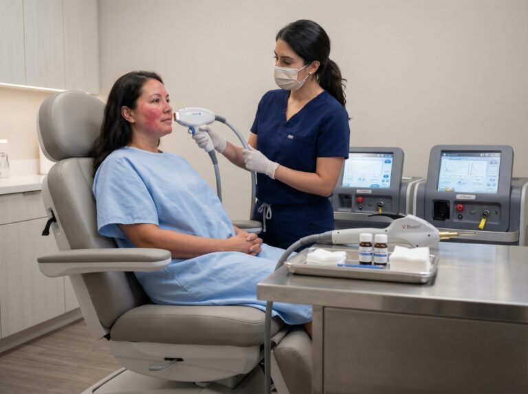
Vascular Dominant
If the circles are blue or purple, vascular lasers (e.g., Nd:YAG, Pulsed Dye) are the solution. Creams with caffeine offer temporary constriction.
| Sign | Likely Cause | Primary Non-Surgical Option |
|---|---|---|
| Shadow disappears with light/flash | Tear Trough / Volume Loss | Dermal Fillers |
| Darkness persists with skin stretch | Hyperpigmentation | Chemical Peels / Picosecond Laser |
| Purple/Blue tint, visible vessels | Vascular / Thin Skin | Vascular Laser / RF Microneedling |
| Puffiness worse in morning | Fluid Retention / Allergies | Lymphatic Massage / Antihistamines |
| Constant bulge, unaffected by sleep | Orbital Fat Prolapse | RF Tightening (Mild) or Surgery (Severe) |
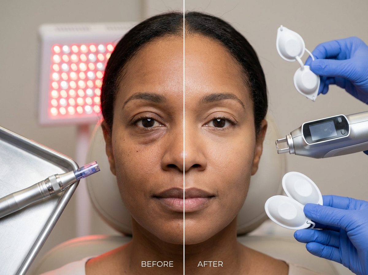
How non-invasive technologies work and what they treat
Understanding biological mechanisms is essential for predicting outcomes. In 2025, treating the periocular area requires specific wavelengths and energy delivery systems designed to respect the 0.5–1.0 mm skin thickness.
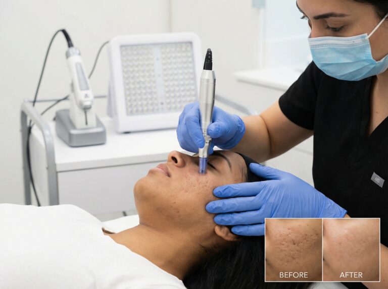
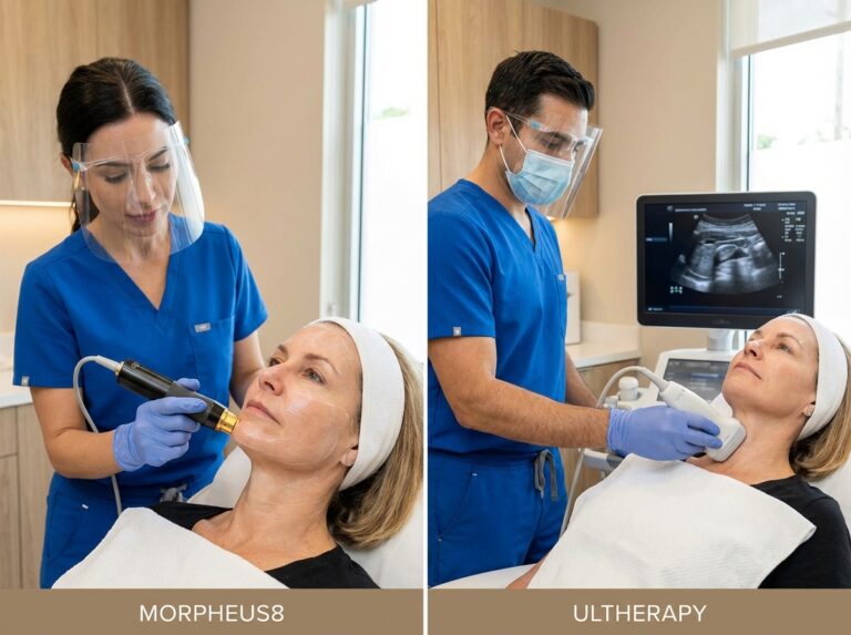
Microneedling and Radiofrequency (RF) Microneedling
Microneedling creates controlled micro-injuries, triggering a wound-healing cascade that releases growth factors and stimulates fibroblasts to produce collagen (neocollagenesis) and elastin.
Manual Microneedling
Used with shallow needle depths (0.25 mm to 0.5 mm) for textural issues like fine lines and crepey skin. It thickens the epidermis to camouflage underlying dark vessels but does not tighten muscle or reduce fat.
RF Microneedling
Insulated needles deliver radiofrequency energy into the dermis, heating tissue to 50–70°C for collagen contraction and remodeling.
* **Indications:** Crepey skin, mild laxity, static wrinkles.
* **Periocular Settings:** Depths limited to 0.5–1.0 mm to avoid orbital septum injury. Energy levels (10–15 J/cm²) are lower than cheek settings to prevent fat necrosis.
* **Pain Level:** Moderate (2–4/10 with topical numbing).
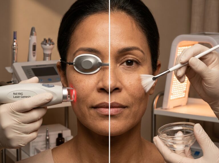
Laser Therapies: Non-Ablative and Picosecond
Non-Ablative Fractional Lasers (1550 nm / 1927 nm)
These devices send columns of heat into the skin while leaving the surface intact. 1550 nm targets water for deep collagen remodeling; 1927 nm targets superficial pigment.
* **Results:** Gradual thickening of the dermis over 3–6 months.
* **Sensation:** Skin may feel like sandpaper (micro-crusting) for 3–5 days post-treatment.
Picosecond Lasers (1064 nm / 755 nm / 532 nm)
The gold standard for pigmented dark circles. Picosecond lasers deliver energy in trillionths of a second, creating a photoacoustic effect that shatters melanin into dust-like particles without burning surrounding tissue.
* **Safety:** Safer for Fitzpatrick skin types IV–VI than thermal lasers, with significantly lower risk of rebound hyperpigmentation.
Superficial Chemical Peels
Peels in the eye area must be superficial, using agents to dissolve desmosomes and lift excess pigment.
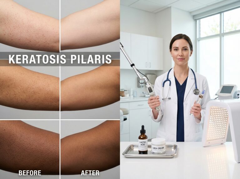
* **Mandelic Acid:** Large molecule, penetrates slowly; ideal for sensitive eye skin and darker tones.
* **Lactic Acid:** Hydrating and gentle.
* **TCA (10–15%):** Used cautiously for texture; higher strengths carry scarring risks in this zone.
LED Phototherapy
Non-thermal photobiomodulation boosts cellular ATP production.
* **Red Light (630–660 nm):** Stimulates collagen and reduces inflammation.
* **Near-Infrared (830 nm):** Improves circulation and reduces cellular inflammation.
* **Role:** Essential adjunct to reduce downtime and enhance healing.
Topical Adjuncts
* **Retinoids:** Low concentrations increase epidermal thickness.
* **Tyrosinase Inhibitors:** Vitamin C, kojic acid, and cysteamine block melanin production. Critical for managing pigmented circles given restrictions on OTC hydroquinone.
* **Caffeine:** Vasoconstrictor for temporary reduction of fluid retention and vascular visibility.
Safety and Periocular Adjustments
Eye Protection
For any light-based treatment inside the orbital rim, **internal corneal shields are mandatory**. These metal contact lenses protect the retina and cornea. External goggles are insufficient for treating the eyelid itself.
Fitzpatrick Skin Typing
Botox and dermal fillers are two of the most effective solutions for under-eye bags and dark circles caused by volume loss and muscle movement, but energy devices require strict adjustment for darker skin (Fitzpatrick IV–VI).
* **Adjustment:** Use lower fluence, longer pulse durations, or non-thermal options (picosecond/microneedling).
* **Pre-treatment:** Darker skin often requires 2–4 weeks of tyrosinase inhibitors before heat-based treatments to suppress melanocytes.
Summary of Modalities and Expectations
| Modality | Primary Target | Typical Sessions | Downtime | Results Timeline |
|---|---|---|---|---|
| RF Microneedling | Laxity, Texture | 3–4 | 2–5 days (swelling) | Gradual (3–6 months) |
| Picosecond Laser | Pigmentation | 2–6 | 1–2 days (redness) | Visible in 4 weeks |
| Non-Ablative Laser | Fine Lines, Texture | 2–4 | 3–5 days (edema) | Gradual (3 months) |
| Chemical Peels | Superficial Pigment | 4–6 | 2–4 days (flaking) | Immediate glow, cumulative |
| LED Therapy | Inflammation, Healing | 6–12 | None | Subtle, supportive |
Selecting and sequencing treatments with practical protocols
The most common failure in treating the under-eye area comes from mismatching the modality to the anatomy. We use a decision framework to match the primary defect with the most effective first-line therapy.
Treatment Selection Algorithm
| Primary Diagnosis | Physical Signs | First-Line Non-Surgical Option | When to Escalate |
|---|---|---|---|
| Fat Prolapse | Puffiness worsens looking up; constant bulge. | RF Microneedling (tightens septum/skin) | Bulge >2 mm requires lower blepharoplasty. |
| Volume Loss (Hollowing) | Shadow disappears with direct lighting. | Collagen induction (Laser/Microneedling) | Deep tear troughs >3 mm need dermal fillers. |
| Pigmentation | Brown discoloration visible in all light. | Picosecond Laser or Chemical Peels | No response after 3 sessions suggests mixed etiology. |
| Vascular | Blue/purple tint or visible vessels. | Vascular Laser (595 nm) or Nd:YAG | Persistent veins may need sclerotherapy (rarely). |
| Skin Laxity | Crepey texture, snap-back test >2 sec. | Fractional Non-Ablative Laser | Ectropion risk or excess skin requires surgery. |
Protocol 1: Microneedling and RF Microneedling
**Ideal Patient:** Fine lines, mild skin laxity, or early-stage fat pad protrusion. Effective for all Fitzpatrick types.
**Preparation:** Stop retinoids 2 weeks prior. Antiviral prophylaxis for patients with a history of cold sores. Avoid isotretinoin for 6–12 months prior.
**Procedure:**
* **Anesthesia:** Topical lidocaine 5% for 45–60 minutes.
* **Depth:** 0.5 mm to 1.0 mm.
* **Energy:** 10–15 J/cm² (lower than face settings).
* **Technique:** 2–3 passes with 10% overlap; pull skin taut.
* **Sessions:** 3–6 sessions spaced 4–6 weeks apart.
**Post-Care:** Erythema and swelling for 24–72 hours; edema up to 5 days. Cold compresses hourly on day one. Avoid active ingredients for 5 days.
Protocol 2: Microneedling with PRP
**Adjustments:** Platelet-Rich Plasma (PRP) is applied topically during and after microneedling. Growth factors penetrate micro-channels, yielding approximately 25% better collagen density than microneedling alone. Superior for thin, transparent skin.
Protocol 3: Non-Ablative Fractional Laser
**Ideal Patient:** Moderate wrinkles and thin skin; cannot afford ablative downtime.
**Procedure:**
* **Safety:** **Metal corneal shields are non-negotiable.**
* **Settings:** Reduce fluence by 30% compared to cheek settings.
* **Sessions:** 2–4 treatments spaced 4 weeks apart.
**Management:** Swelling peaks at 48 hours. Skin feels like sandpaper for 3–5 days. Maintain moisture with bland ointments.
Protocol 4: Pigment Correction (Picosecond and Peels)
**Picosecond/Q-Switched Laser:** Low-fluence 1064 nm.
* **Endpoint:** Mild erythema only. No frosting.
* **Sessions:** 2–6 sessions every 4 weeks.
**Superficial Chemical Peels:** Mandelic, Lactic, or low-strength TCA (3%–6%).
* **Application:** Semi-dry q-tip, 2 mm from lash line.
* **Neutralization:** Immediate if erythema appears.
* **Frequency:** 4–6 peels every 2–3 weeks.
Protocol 5: LED Phototherapy
**Protocol:** Red (633 nm) and Near-Infrared (830 nm).
* **Timing:** Immediately following microneedling or laser for 20 minutes.
Sequencing and Combination Treatments
1. **Surface to Deep:** Address surface pigmentation first if using lasers absorbed by surface melanin.
2. **Tighten then Fill:** Perform energy treatments (Laser/RF) to thicken the dermis first. Wait 4–8 weeks before placing dermal fillers. A thicker dermis hides the filler better and reduces the Tyndall effect (blue cast).
3. **Botox Timing:** Neuromodulators can be done the same day if placed *after* energy treatments. If done before, wait 2 weeks to avoid product displacement.
**Sample Timeline:**
* Month 1: Picosecond laser + LED.
* Month 2: RF Microneedling.
* Month 3: RF Microneedling + PRP.
* Month 5: Assessment for Dermal Fillers.
Contraindications and Safety Precautions
* **Absolute Contraindications:** Pregnancy/breastfeeding, active infection (Herpes/bacterial), recent isotretinoin use (within 6–12 months), unrealistic expectations.
* **Specific Precautions:**
* **Eye Protection:** Internal metal corneal shields are required for lasers inside the orbital rim.
* **Skin Type:** For Fitzpatrick IV–VI, reduce energy by 50% and extend intervals to minimize PIH.
* **Vascular Limits:** Vascular lasers can cause bruising lasting 7–10 days (“social downtime”).
Cost Expectations and Maintenance
Insurance typically does not cover these cosmetic procedures. Costs reflect 2025 pricing in the United States.
| Treatment Modality | Cost Per Session (USD) | Typical Series Cost (USD) | Maintenance Schedule |
|---|---|---|---|
| Microneedling (Standard) | $300 – $600 | $1,500 (4 sessions) | Every 6 months |
| RF Microneedling | $800 – $1,500 | $3,500 (3 sessions) | Once yearly |
| Non-Ablative Laser | $500 – $1,200 | $2,500 (3 sessions) | Once yearly |
| Chemical Peel (Eye) | $150 – $300 | $800 (4 sessions) | Quarterly |
| Picosecond Laser | $400 – $800 | $2,000 (4 sessions) | As needed |
Frequently Asked Questions
What are the risks and how do we minimize them?
The periocular area carries specific risks. Post-inflammatory hyperpigmentation (PIH) is the main concern for darker skin types (Fitzpatrick IV-VI). We minimize this by using lower energy settings, longer intervals, and priming with tyrosinase inhibitors. Eye safety is critical; lasers used inside the orbital rim require internal metal corneal shields to protect the retina. **Patient Action Item:** Ask your provider explicitly if they use metal eye shields. If they say shields are not necessary for work inside the bone rim, seek a second opinion.
Can treatments be combined in a single visit?
Stacking treatments is effective but requires caution. We often combine a chemical peel with LED therapy in the same visit to reduce inflammation. However, we rarely combine aggressive heat-based treatments with injectables on the same day. Heat can degrade fillers or increase neurotoxin diffusion. A safe sequence is energy devices first, followed by fillers 4 weeks later.
What should I avoid before and after procedures?
Avoid blood thinners (aspirin, ibuprofen, fish oil, Vitamin E) for 7 days prior to minimize bruising. Stop retinoids and active acids 5–7 days before treatment. Post-procedure, treat the skin like a fresh wound: avoid direct sun, saunas, hot yoga, and swimming for at least 7 days. Use physical sunscreens (zinc/titanium) rather than chemical ones.
Who is not a candidate for these treatments?
Patients with active infections (cold sores, conjunctivitis), pregnant or breastfeeding women, and those with history of keloid scarring are generally excluded. Anatomically, patients with significant ectropion (lower lid laxity) or massive fat herniation are surgical candidates; non-invasive devices will not provide satisfactory results.
Are at-home devices effective?
Home beauty tech (LED masks, microcurrent) is safe but significantly less powerful than clinical equipment. A professional RF device might reach 60°C in the dermis; home devices cap at 40–42°C. They are maintenance tools, not corrective ones. The Under Eye Patches Market is growing because these products offer hydration and temporary depuffing, but they do not alter skin structure.
Is there any insurance coverage?
Treatment for under-eye bags and dark circles is considered cosmetic and is an out-of-pocket expense. The only exception usually involves upper eyelid surgery (blepharoplasty) if sagging skin obstructs vision. HSA/FSA funds are typically not eligible unless there is a specific medical diagnosis like trauma scarring.
Conclusions and practical takeaways
We have explored the mechanisms of microneedling, lasers, and peels. The technology available in late 2025 offers impressive tools, but success relies on selecting the right candidate and managing the biological reality of the 0.5 mm thick periorbital skin.
Surgery Versus Non-Surgical Interventions
We must be honest about limitations. Energy devices improve skin quality, thicken the dermis, and reduce pigmentation, but they do not remove structural fat. If a patient presents with significant fat prolapse (>2 mm), surgery remains the gold standard. Attempting to camouflage a large fat pad with filler often leads to an overfilled, puffy appearance. Non-surgical options shine for patients under 40 with mild hollowing or textural issues, where the goal is preservation and subtle correction.
Tailoring Treatment to Skin Type
For Fitzpatrick skin types IV through VI, safety is prioritized over speed. Picosecond lasers are preferred for pigment because they rely on acoustic energy rather than heat, reducing the risk of PIH (which stands at roughly 40% if settings are incorrect). Mandelic acid peels are a safer chemical alternative. Aggressive fractional CO2 lasers are generally avoided in this area for darker skin due to scarring risks.
Realistic Expectations and Maintenance
Collagen synthesis takes time. While immediate tightening from edema may occur, real structural improvement peaks around three months post-treatment. Maintenance is mandatory; aging continues, and vessels may re-dilate.
| Primary Concern | Preferred Modality | Typical Sessions | Time to Full Result | Maintenance |
|---|---|---|---|---|
| Volume Loss (Hollowing) | Dermal Fillers / PRP | 1-2 | 2 weeks | 6-12 months |
| Pigmentation (Brown) | Picosecond Laser / Peels | 3-6 | 8-12 weeks | Daily SPF + Yearly |
| Vascular (Blue/Purple) | Pulsed Dye Laser / Nd:YAG | 2-4 | 4-8 weeks | 6-12 months |
| Texture / Crepey Skin | RF Microneedling / LED | 3-4 | 3-6 months | Yearly |
The Patient Checklist
The global eye cream market size is expected to reach $5.3 billion in 2025, indicating massive demand, but products have limits. Before booking professional treatment:
* **Identify the Root Cause:** Use the lighting and stretch tests described earlier.
* **Check Credentials:** Ensure the provider has specific training in eye anatomy and uses internal corneal shields for lasers.
* **Discuss Skin Type:** Explicitly ask about PIH mitigation for darker skin tones.
* **Plan for Downtime:** Do not book immediately before major events.
* **Understand Costs:** Budget for a full series ($1,500–$5,000), as insurance does not cover these procedures.
Forward-Looking Remarks
As we move through late 2025, the trend is toward combination therapies—stacking modalities like peels and LED to reduce downtime and enhance results. Hybrid devices combining picosecond domains with radiofrequency are emerging, promising to target pigment and laxity simultaneously.
Safety First: The eye is a sensory organ critical for life quality. No cosmetic improvement is worth risking vision. Corneal shields are mandatory for laser procedures around the eye. Conservative treatment planning is always the smarter path.
References
- Under Eye Patches Market – Future Market Insights — The Global Under-Eye Patches Market is projected to grow from USD 817.8 million in 2025 to USD 1,686.7 million by 2035, at a CAGR of 7.51%.
- Comprehensive Overview of Dark Circles Removing Eye Cream … — The global market for dark circles removing eye cream is experiencing robust growth, projected to reach an estimated $5,500 million by 2025, …
- Eye Creams For Dark Circles Market Report – Dataintelo — The global market size for eye creams targeting dark circles was valued at USD 2.1 billion in 2023 and is forecasted to reach approximately USD 4.5 billion …
- Under Eye Serum Market Size, Share and Analysis, 2025-2032 — The global under eye serum market is estimated to be valued at USD 4.83 Bn in 2025 and is expected to reach USD 11.22 Bn by 2032, exhibiting a compound annual …
- Dark Circle Treatments in 2025 – The Skin Company — Botox and dermal fillers are two of the most effective solutions for under-eye bags and dark circles caused by volume loss and muscle movement.
- Under Eye Bags Makeup Trend: Why Concealers Are Out in 2025 — The peak in January 2025 with a value of 88 is notable. "Eye bag patches" also started showing up in August 2024, which might indicate a growing …
- eye fillers for dark circles: 7 Powerful Benefits in 2025 — Discover eye fillers for dark circles, how they work, who's eligible, benefits, costs, recovery tips, risks, and expert guidance.
- Eye Cream Market Size, Share & Analysis Report, 2025-2034 — The global eye cream market size was estimated at USD 5 billion in 2024. The market is expected to grow from USD 5.3 billion in 2025 to USD 9.5 billion in 2034 …
- Clinical Efficacy of a Novel Topical Formulation on Periorbital Dark … — Hyperpigmentation and periorbital dark circles remain challenging dermatological concerns due to their multifactorial etiology, …